iFluor - Ultrawide FOV imaging
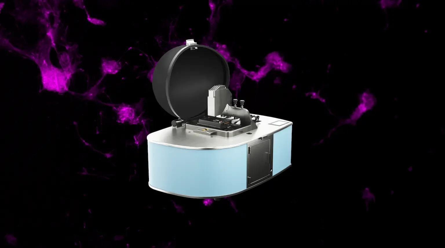
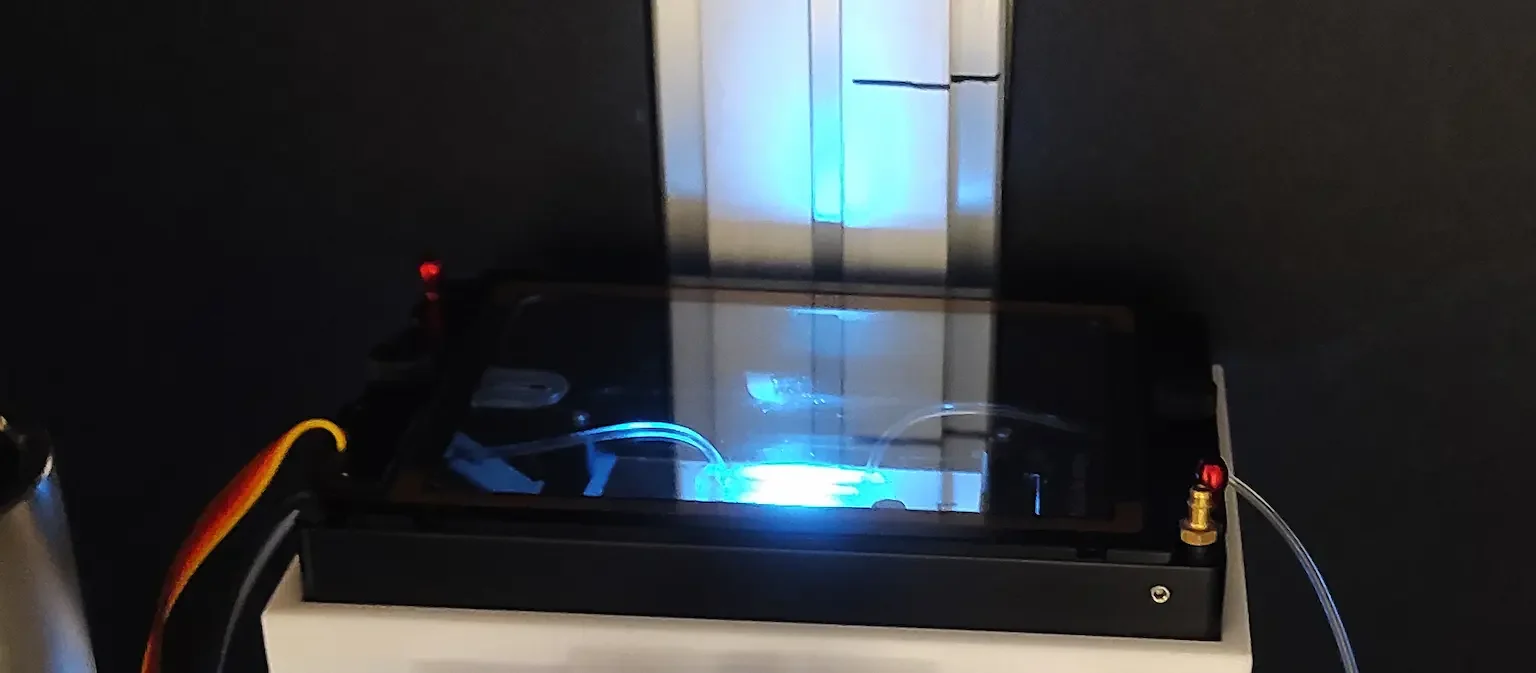
The iFLUOR integrates advanced microfluidics with a 10×10 mm high‑resolution imaging field, allowing cells to be imaged continuously without moving the sample and thereby minimizing disturbances and the risk of bubble formation. An onboard leak detector provides instant—optionally remote—alerts to ensure the microfluidic circuit operates reliably, safely, and with consistent performance.
-
Ultrawide Field of View
Capture large populations of cells or organisms with a wide 10x10 mm imaging area at high resolution, ideal for statistical analysis and large-scale experiments.
-
Versatile Imaging
Supports both transmission imaging and single-color fluorescence, enabling researchers to view the same sample in multiple ways without moving the sample.
-
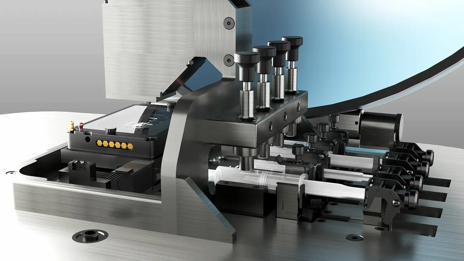
Advanced Microfluidics
Up to 4 integrated microfluidic channels with programmable fluidic delivery for real-time control of the environment, perfect for dynamic, long-term cell experiments.
-
Enhanced Adaptability
Compatible with customizable chip designs for specific research needs, such as barrier-controlled environments for small organisms like C. elegans.
-
All-in-one Software
The all-in-one software allows for timelapse design, automated image acquisition, data analysis, and control over microfluidic delivery, simplifying complex experiments.
Applications
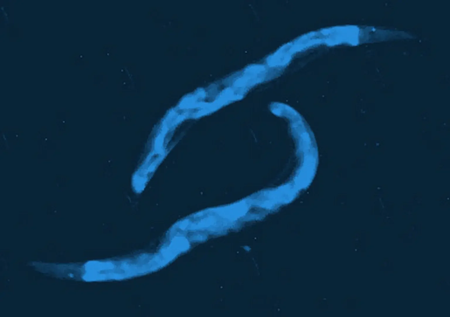
C. elegans
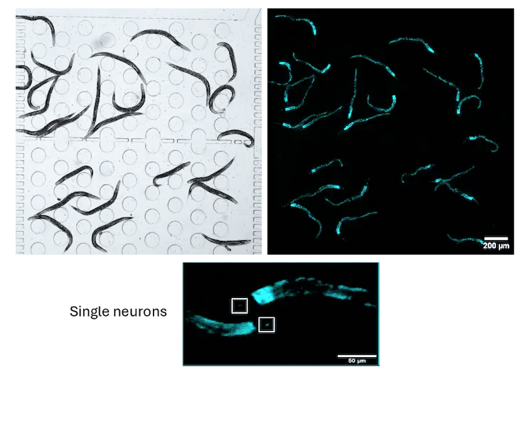
Ultra-wide field acquisition of a population of C. elegans in a microfluidic chip.
Transmission channel (left) and fluorescence channel (right) images of C. elegans with calcium indicator expression in a microfluidic chip. Scale bar 200 µm. Zoomed-in image shows single neurons with calcium indicator fluorescence in the head of C. elegans. Scale bar 50 µm.
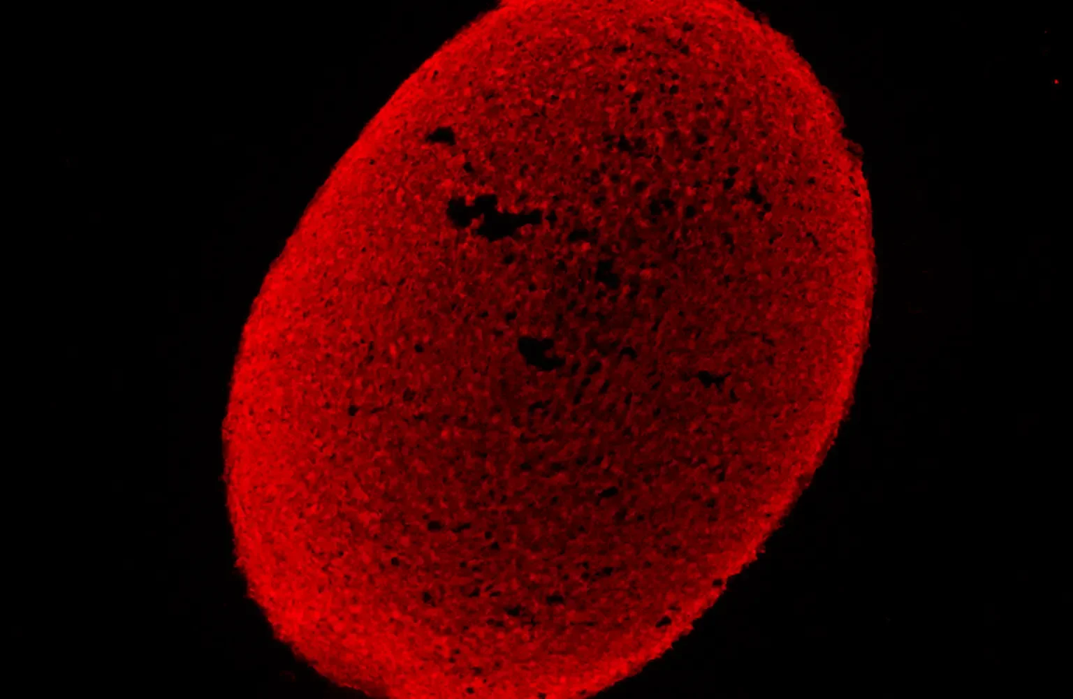
Neural rosettes
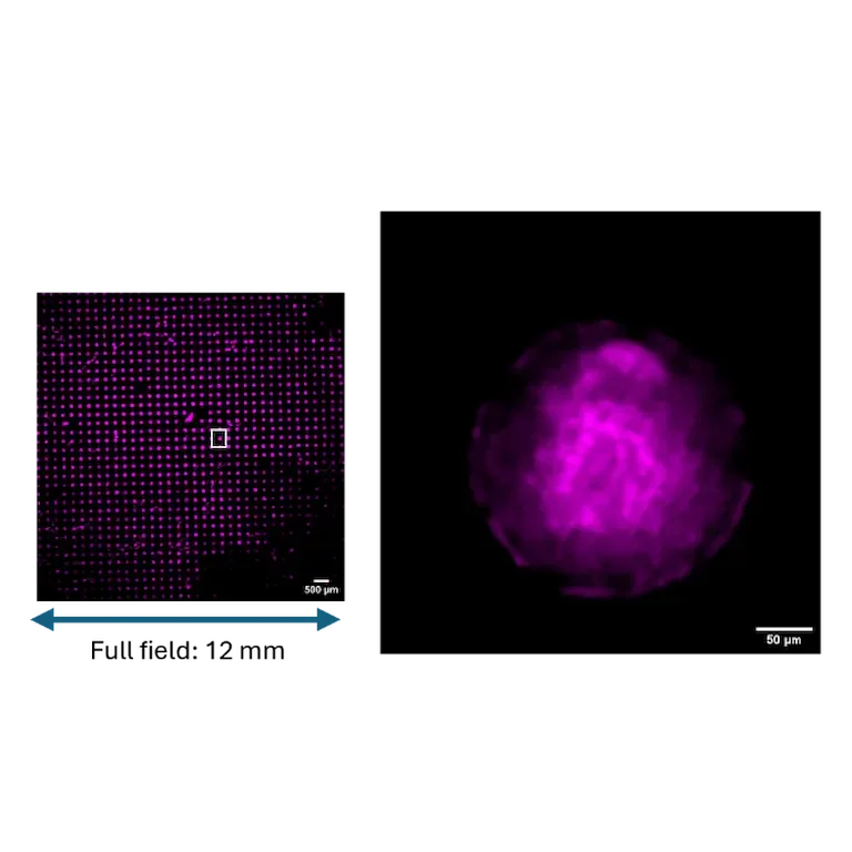
Ultra-wide field acquisition of hundreds of human iPSC-derived neural rosettes on micropatterned support.
Representative images of full-field acquisition (left) of neural rosettes immunostained with Atto 532-Phalloidin (Sigma Aldrich). Scale bar 500 µm (n= 900 rosettes). Zoomed-in and super-resolved image (right) shows a single neural rosette. Scale bar 50 µm.
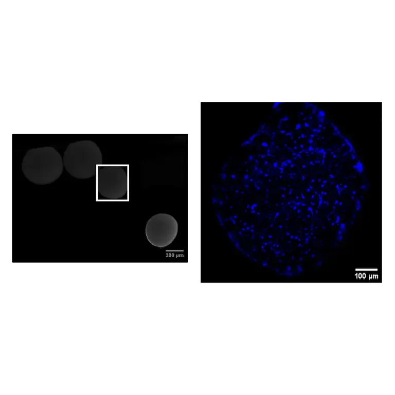
Organoids
iFLUOR's advanced microfluidics system facilitates the growth, maintenance, and imaging of organoids under controlled conditions
Acquisition of cortical organoids with anti-Pax6 antibody (Santa Cruz Biotechnology) (left). Zoomed-in image (right) shows a single cortical organoid. Scale bar 100 µm. Researchers can simulate and monitor development in real-time, providing critical insights into organ formation and disease modeling.